.png)
Pleuroscopy, also known as medical thoracoscopy, is an advanced minimally invasive procedure that allows direct visualisation of the pleural cavity — the space between the lungs and the chest wall — using a thin camera-equipped instrument called a thoracoscope.
Unlike CT scans and X-rays that provide images from outside the body, pleuroscopy allows direct visualisation of the pleura and enables targeted biopsies from abnormal areas, making it one of the most valuable procedures for diagnosing unexplained pleural diseases.
At Healthy Chest & Sleep Clinic, Dr. Vishal Sharma offers pleuroscopy / thoracoscopy for the diagnosis and management of complex pleural disorders without the need for major surgery in many cases.
Book a consultation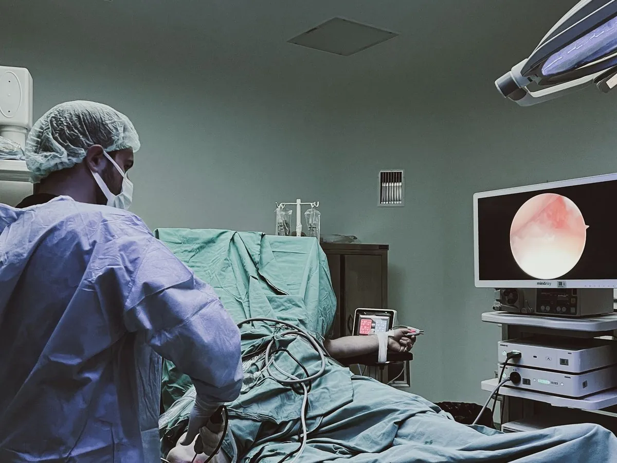
Your pulmonologist may recommend pleuroscopy when routine investigations cannot determine the cause of a pleural problem.
Fluid repeatedly accumulates despite treatment
Cause unclear after fluid analysis and imaging
TB suspected but not confirmed by routine tests
To obtain tissue for a definitive diagnosis
Abnormal thickening seen on CT scans
Breathlessness, chest pain or unexplained findings
Pleuroscopy is frequently used in the diagnosis of a wide range of pleural diseases that require direct visualisation and biopsy.
One of the most common causes of unexplained pleural effusion in India.
Including metastatic cancers affecting the pleura.
A rare cancer involving the pleural lining.
Persistent inflammation requiring tissue diagnosis.
When the underlying cause remains uncertain.
Abnormal findings requiring biopsy and evaluation.
In the past, tissue samples were often obtained through blind biopsy techniques. These procedures had limitations because the doctor could not directly see the abnormal area.
Pleuroscopy allows direct visualisation, targeted biopsy, higher diagnostic accuracy, better tissue sampling and improved treatment planning — significantly improving the likelihood of obtaining a definitive diagnosis.
Pleuroscopy follows a careful, structured process designed to ensure comfort, safety and an accurate diagnosis.
Symptoms, CT findings, X-rays, pleural fluid reports, medical and medication history are reviewed.
The procedure is typically performed under sedation and local anaesthesia, prioritising comfort and safety.
A small opening is created in the chest wall to introduce the thoracoscope into the pleural cavity.
The pleural cavity is examined for inflammation, nodules, thickening, infection and abnormal tissue.
Samples are obtained from abnormal areas for histopathology, TB testing and cancer diagnosis.
Pleuroscopy is not only a diagnostic procedure. In selected patients, it can also be used therapeutically.
Large pleural effusions causing breathlessness can be drained.
A procedure performed to prevent recurrent fluid accumulation in certain patients.
Reducing fluid often leads to immediate improvement in breathing comfort.
Pleuroscopy is considered safe when performed by trained specialists. Before recommending it, Dr. Vishal Sharma carefully evaluates every relevant factor.
A complete review of your general wellbeing.
Checking how well your lungs are working.
Studying scans to plan a targeted approach.
Assessing conditions that may affect the procedure.
Every effort is made to minimise risk and ensure safety.
Recovery varies depending on the individual and the nature of the procedure. Detailed post-procedure instructions are provided to support a smooth recovery.
Most patients tolerate the procedure well.
Noticeable relief if fluid has been drained.
A brief stay for most patients.
A swift return to normal daily life.
Specialised training and experience in advanced pleural procedures.
Accurate diagnoses while minimising patient discomfort.
Diagnosis, treatment planning and follow-up under one roof.
Every recommendation is tailored to the patient's condition.
Combining advanced technology with evidence-based care.
Pleuroscopy provides a highly accurate, minimally invasive approach to evaluating pleural abnormalities, recurrent fluid collections, tuberculosis and pleural cancers. Consult Dr. Vishal Sharma to determine whether pleuroscopy / thoracoscopy is appropriate for your condition.